Another day, another personal health-themed news post. Wait, didn't I just say that less than two weeks ago? This time, however, I'm not talking about directly testing a human subject, but instead evaluating what he or she is about to ingest. And the topic has personal relevance; a friend's mother was sickened (but thankfully recovered) five years ago due to the E. coli outbreak in spinach. But unfortunately, this particular piece of equipment wouldn't have helped her unless she had first put the spinach in a blender and added water…since it only works on liquids.
As discussed at The Verge (and later also picked up by Gizmodo), researchers at UCLA have developed a cameraphone accessory that calculates the concentration of E. coli in a sample. From The Verge's writeup:
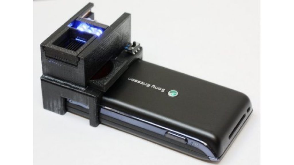
The device works similarly to a fluorescence microscope, pumping the sample into a series of small tubes treated with E. coli antibodies, then measuring the excitement of quantum dots (small fragments of semiconductors) placed around them with the phone's camera and an additional lens. From this excitement, researchers can calculate the concentration of E. coli in the sample. The method was tested successfully both in a specially prepared solution and in a glass of milk.
Ozcan's research group has been working for some time on scaling down biomedical technologies to fit on phones. Last year, they created a flow cytometer, a machine used to image cells and bacteria that could be built for five dollars and attached to a cell phone. This latest attachment builds on the cytometer, but tests for specific pathogens rather than white blood cells, the focus of the previous study.
And for those of you who, like me, aren't offhand familiar with the function of a fluorescence microscope, here's Wikipedia's sound bite summary:
A sample is illuminated with light of a wavelength which causes fluorescence in the sample. The light emitted by fluorescence, which is at a different, longer, wavelength than the illumination, is then detected through a microscope objective. Two filters are normally used in this technique; an illumination (or excitation) filter which ensures the illumination is near monochromatic and at the correct wavelength, and a second emission (or detection) filter which ensures none of the excitation light source reaches the detector.
For more on cameraphone (and more general smartphone) uses in personal healthcare, check out this Slashdot highlight from yesterday, which links to a New York Times writeup.